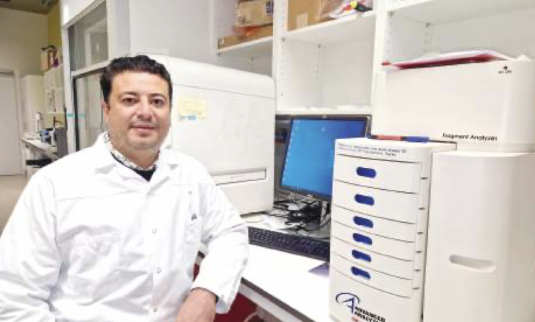
In a significant breakthrough for the field of genomics, a leading researcher based at the University of Geneva, Egyptian scientist Haitham Shaaban, has unveiled new insights into the genetic origins of cancer and aging.
Shaaban is a specialist in developing digital microscopy to monitor the instantaneous interactions of genetic material within cells.
This aligns with the promising field of genomics, the future of medicine in coming decades.
It is one of the most important mechanisms for early disease diagnosis, relying on applications of artificial intelligence and genetics.
This approach accurately identifies diseases, determines the interaction with patients, and identifies appropriate dosages for each treatment, opening the door to saving millions of lives through early detection of diseases, including cancer and aging.
His findings, published in the world-renowned journal Nature, offer a more refined understanding of the complex interplay between genetic sequences and proteins.
This groundbreaking research marks a pivotal moment in the quest to combat these age-old diseases.
In an exclusive interview with Al-Masry Al-Youm, the Egyptian genomics expert revealed that genome regulation is an exceedingly intricate process. It involves countless proteins interacting with genetic material at lightning speed, rendering the genetic structures highly dynamic and mobile enough to sustain cellular function.
As Shaaban explained, in order to fully grasp the intricacies of genome regulation in healthy cells and the disruptions that occur in diseased cells like cancer and aging cells, it is vital to scrutinize the nuclear protein interactions with genetic material using high-resolution techniques capable of visualizing these interactions in living cells.
He explained that “We’ve recently developed a new microscopy technique, Hi-D mapping, to visualize how our genome is organized. This has allowed us to study the dynamics of genetic material. While promising, the initial version had limitations, including the inability to study protein interactions and a reliance on powerful computers and skilled researchers.”
The Egyptian scientist confirmed in this study, published in Nature Protocols, they developed a computational optical microscopy technique to overcome these limitations and visualize and study the functional organization of the genome, as well as analyze interacting proteins such as histones (H2B) and various ribonucleic acids (RNA) with DNA sequences in real time.
This technique integrates optical flow algorithms, Bayesian statistics, and machine learning frameworks for the probabilistic modeling of spatial distributions of DNA sequences, H2B, and RNA in both healthy and cancerous cells using various optical microscopy methods, he added.
According to Shaaban, this novel technique enabled his team to accurately detect the dynamics of diffusion and biophysical information, and to model physical diffusion from data with high spatial resolution for the entire genome in living cells.
This allows for the classification and mapping of these cells in less than an hour of analysis time.
Furthermore, this technique allows for the observation of the spatiotemporal dynamics within live cell nuclei, aiding in deciphering the dynamic linkage of gene expression to higher-order chromatin structures at the genome level in human cells.
The Egyptian genomics scientist revealed that this technique can potentially be applied to study tissues and examine various cell types, providing an approach to investigate the diverse regulation of genomics to understand cellular functions and study diseases linked to genome regulation.
Additionally, this technique will enable researchers to explore the complex dynamics of the genome and nuclear proteins in real time, providing valuable insights into gene regulation and cellular function in both health and disease.